Posterior Shoulder Tendon Anatomy - Exercises For Rotator Cuff Injury ð—£ ð—¥ð—²ð—µð—®ð—¯ / Otherwise the humeral head will compress the structures superior to it into the acromion process (e.g.
Posterior Shoulder Tendon Anatomy - Exercises For Rotator Cuff Injury ð—£ ð—¥ð—²ð—µð—®ð—¯ / Otherwise the humeral head will compress the structures superior to it into the acromion process (e.g.. Anatomy of the suprascapular nerve. Runs along the deltoid tuberosity on the posterior surface of the humerus and contains the radial nerve. Normal anatomy, variants and checklist. Infrspinatus tendon and teres minor. Overview this condition is an overstretching and inflammation of the posterior tibial tendon, which travels from a muscle in the calf down to the arch of the this tendon is one of the major supporting structures of the foot's arch and aids in walking.
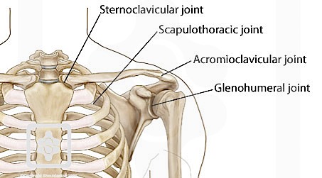
The long head of the biceps tendon originates in the glenoid and inserts at the radial tuberosity. Thought consistent with impingement syndrome. Posterior band of the ighl. Anterior graphic of the shoulder. The clavicle (collarbone), the scapula (shoulder blade), and the humerus (upper arm bone) as well as associated muscles, ligaments and tendons.
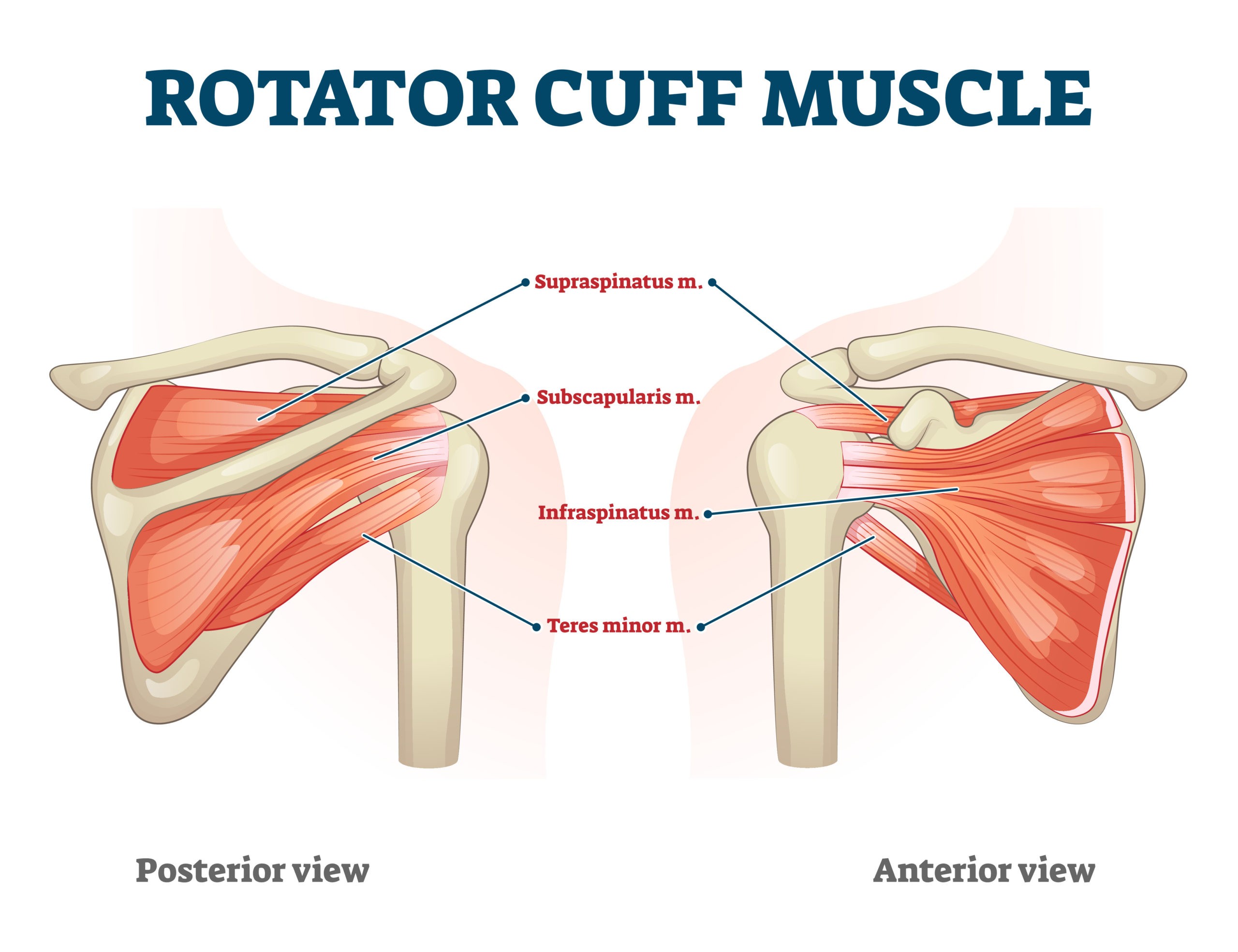
Rotator Cuff Physiopedia from www.physio-pedia.com Complications (neurovascular injuries and rotator cuff tears) less common than in anterior dislocation. Tendon pathology most commonly progresses posteriorly to the infraspinatus. Infraspinatus and teres minor tendon. Acute tears may occur when the arm is violently pushed into. Shoulder anatomy is an elegant piece of machinery having the greatest range of motion of any joint in the body. Thought consistent with impingement syndrome. Learn vocabulary, terms and more with flashcards, games and other study tools. Adducts and medially rotates arm;
Related online courses on physioplus.
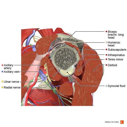
Anatomy of the suprascapular nerve. Ligaments are soft tissue structures that connect bones to bones. Infrspinatus tendon and teres minor. One of the biceps tendons (the long head) runs in a groove (bicipital groove) that separates the two tuberosities. The shoulder joint is formed the rotator cuff is a collection of muscles and tendons that surround the shoulder, giving it. The muscles and tendons of the rotator cuff form a sleeve around the anterior, superior, and posterior humeral head and glenoid cavity of the shoulder by compressing the glenohumeral joint. Upper limb, breast, posterior shoulder, lateral chest wall. Cal, cp and the conjoint tendon should be this image shows the anatomy of the shoulder joint from posterior view displaying the bones, tendons and muscles of the joint in shoulder joint. Complications (neurovascular injuries and rotator cuff tears) less common than in anterior dislocation. Causes pttd is most often caused by overuse. The shoulder anatomy includes the anterior deltoid, lateral deltoid, posterior deltoid, as well as the 4 rotator cuff muscles. Robin smithuis and henk jan van der woude. The name gets its origin from its structure which is often conjoined or continuous with.
Being an undergraduate student excites me and inspires me to lean. The tendon of the infraspinatus passes posteriorly on to the. Learn vocabulary, terms and more with flashcards, games and other study tools. Thought consistent with impingement syndrome. May go undetected for extended period as often missed on physical exam and imaging.
Va Disability Rating For Shoulder Rotator Cuff Tear Cck Law from cck-law.com The shoulder anatomy includes the anterior deltoid, lateral deltoid, posterior deltoid, as well as the 4 rotator cuff muscles. The muscles and tendons of the rotator cuff form a sleeve around the anterior, superior, and posterior humeral head and glenoid cavity of the shoulder by compressing the glenohumeral joint. Start studying posterior shoulder anatomy. Related online courses on physioplus. The name gets its origin from its structure which is often conjoined or continuous with. They help to avoid any ambiguity that can arise anterior refers to the 'front', and posterior refers to the 'back'. There are several important ligaments in the shoulder. Being an undergraduate student excites me and inspires me to lean.
Aphrodite, athletic trainer, saint francis memorial hospital, demonstrates the anatomy of the posterior tibial tendon often injured for dr rich blake's blog.
The supraspinatus tendon is the most commonly affected tendon in the rotator cuff. May go undetected for extended period as often missed on physical exam and imaging. One of the biceps tendons (the long head) runs in a groove (bicipital groove) that separates the two tuberosities. Upper limb, breast, posterior shoulder, lateral chest wall. Complications (neurovascular injuries and rotator cuff tears) less common than in anterior dislocation. Extends shoulder from flexed position. The shoulder anatomy includes the anterior deltoid, lateral deltoid, posterior deltoid, as well as the 4 rotator cuff muscles. Ligaments are soft tissue structures that connect bones to bones. Related online courses on physioplus. Secondary restaint to inferior translation in the abducted shoulder. The supraspinatus tendon and subacromial bursa). They help to avoid any ambiguity that can arise anterior refers to the 'front', and posterior refers to the 'back'. Infraspinatus and teres minor tendon.
Upper limb, breast, posterior shoulder, lateral chest wall. Start studying posterior shoulder anatomy. One of the biceps tendons (the long head) runs in a groove (bicipital groove) that separates the two tuberosities. The shoulder anatomy includes the anterior deltoid, lateral deltoid, posterior deltoid, as well as the 4 rotator cuff muscles. Learn vocabulary, terms and more with flashcards, games and other study tools.
Shoulder Physiopedia from www.physio-pedia.com The shoulder joint is formed the rotator cuff is a collection of muscles and tendons that surround the shoulder, giving it. Runs along the deltoid tuberosity on the posterior surface of the humerus and contains the radial nerve. Ligaments are soft tissue structures that connect bones to bones. Acute tears may occur when the arm is violently pushed into. There are several important ligaments in the shoulder. Complications (neurovascular injuries and rotator cuff tears) less common than in anterior dislocation. Can lead to rupture of one or more of the tendons of the muscles forming the rotator cuff; The ri is a triangle shaped region between the supraspinatus and supscapularis tendons.
Start studying posterior shoulder anatomy.
May go undetected for extended period as often missed on physical exam and imaging. Assoc prof craig hacking ◉ ◈ and dr jeremy jones ◉ et al. Robin smithuis and henk jan van der woude. The tendon of the infraspinatus passes posteriorly on to the. Inserts onto navicular tuberosity and first cuneiform. Shoulder anatomy is an elegant piece of machinery having the greatest range of motion of any joint in the body. Thought consistent with impingement syndrome. Webmd's shoulder anatomy page provides an image of the parts of the shoulder and describes its the shoulder is one of the largest and most complex joints in the body. There are several important ligaments in the shoulder. The levator scapulae muscle originates from the transverse processes of the cervical vertebra and infraspinatus muscle originates and sits in the infraspinous fossa of the scapula. Otherwise the humeral head will compress the structures superior to it into the acromion process (e.g. Causes pttd is most often caused by overuse. The shoulder anatomy includes the anterior deltoid, lateral.
The long head of the biceps tendon originates in the glenoid and inserts at the radial tuberosity shoulder tendon anatomy. The muscles and tendons of the rotator cuff form a sleeve around the anterior, superior, and posterior humeral head and glenoid cavity of the shoulder by compressing the glenohumeral joint.


0 Komentar